health and healing
Advanced imaging offers selected stroke patients a chance beyond 24 hours
Advanced brain imaging is giving some people in Vietnam who arrive late after an ischemic stroke a renewed chance of treatment. CT or MRI perfusion scans can distinguish tissue that is already damaged from tissue that may still recover, helping specialists decide whether clot removal could be worthwhile beyond the conventional treatment window.
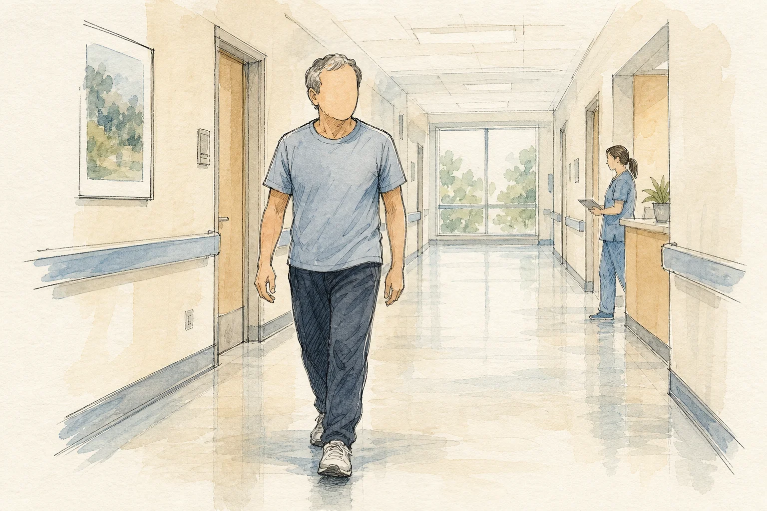
At an August stroke meeting in Ho Chi Minh City, clinicians described a 27-year-old man whose initially mild speech and weakness symptoms deteriorated into paralysis on one side about 60 hours after onset. Perfusion imaging showed that a substantial area of brain tissue could still be saved. After mechanical thrombectomy, he recovered quickly and was walking almost normally around 10 days later.
The case reflects emerging evidence, not a blanket extension of treatment. A Vietnamese study of patients arriving more than 24 hours after onset found that about half could walk nearly independently after three months. Findings presented from the LATE-MT study also indicated that carefully selected patients may benefit from thrombectomy between 24 and 72 hours when imaging identifies recoverable tissue.
The potential is especially relevant in Vietnam, where clinicians at a major stroke center reported that only 12% of patients arrived within three hours. But the expanded window does not make waiting safe. Treatment after 24 hours requires strict assessment and carries serious risks, so sudden facial drooping, limb weakness, speech difficulty or loss of balance still demands immediate emergency care.