technology and innovation
Fluorescent probes bring DNA folding into sharper view inside living cells
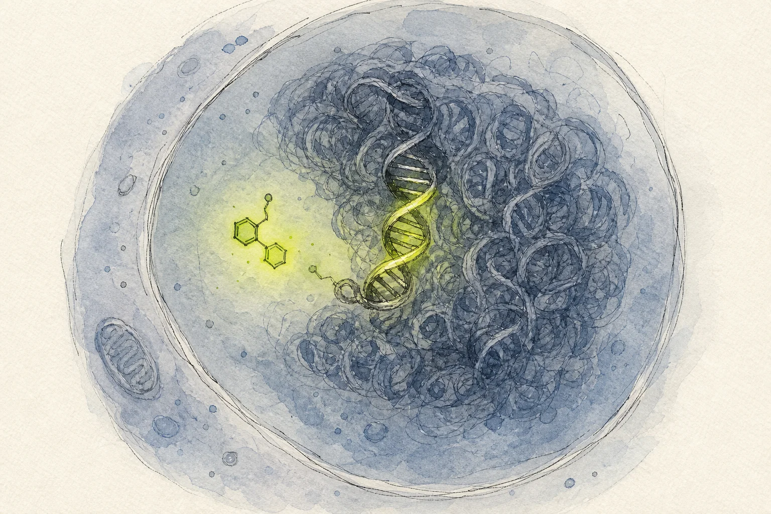
Researchers led by Barcelona’s Centre for Genomic Regulation have developed fluorescent probes that reveal how DNA is packed and moves inside living cells. The molecules, called HoTs, produced images at 20-nanometre resolution in live cells and located individual dye molecules with three-nanometre precision in preserved laboratory cells, close to the width of DNA itself.
The probes cross cell membranes, attach to DNA and switch between bright and dark states on their own. Advanced microscopes can pinpoint the blinking molecules separately, then computationally combine their positions into a detailed image. In living cells, the method avoids the harsh chemicals and intense laser light required by many existing single-molecule techniques.
When applied to intestinal tissue sections from three people with cancer, the probes showed that DNA in tumour cells was looser and more dispersed than in neighbouring healthy tissue. The finding was consistent with earlier research, although the study did not establish why this change occurs or validate it as a diagnostic measure.
In a separate test, an artificial intelligence system distinguished live skin cells from stem cells with 96 to 98 per cent accuracy by analysing their differently folded DNA. The probes label the whole genome rather than particular genes, but the work, published in Molecular Cell, offers a new way to study chromatin architecture in motion.